DIAGNOSIS

Knowing if a person is having a seizure and diagnosing the type of seizure or epilepsy syndrome can be difficult. There are many other disorders that can cause changes in behaviour and can be confused with epilepsy. Since the treatment of seizures depends on an accurate diagnosis, making sure that a person has epilepsy and knowing what kind is a critical first step.

Neurological examination is a key step in the diagnosis.

Clinical history and neurological examination are the basis for the diagnosis of seizures and, consequently of epilepsy. Often the doctor asks for a laboratory evaluation (tests), but they are considered as additional diagnosis tests. Blood tests are used to check your global Health status and to screen for any other medical condition that may be the cause of epilepsy.

The history of the characteristics of the disease is the first important step in diagnosis.

The history of the characteristics of the disease is the first important step in the diagnosis. The doctor needs all the information about what happened before, during, and after your seizures, so he will probably ask about the premonitory signs and the details of the seizure (how it happened, duration, feelings and sensations before, during and after the epileptic seizure). If you cannot give enough information, then others who have seen the seizures happen should contribute what they know.

After knowing the person’s clinical history, neurological examination is a key step in the diagnosis, being able to identify or localize cerebral pathologies. The general physical examination is also important to determine if the person has any underlying condition. For example, abnormal skin markings could indicate a neurocutaneous disorder in which epilepsy is common, such as tuberous sclerosis or neurofibromatosis. 
 
Even with accurate descriptions of the events, sometimes other tests are needed to learn more about the brain, what is causing the events, and where the problem is located. The most common tests are an electroencephalogram (EEG) test and a magnetic resonance imaging (MRI).
 

EEG | ELECTROENCEPHALOGRAM

An electroencephalogram (EEG) is a test to detect abnormalities in the electrical activity of the brain. Brain cells (or neurons) communicate by producing electrical signals. To perform an EEG test, electrodes are placed on the scalp to detect and record patterns of this electrical activity and check for abnormalities, such as focal points or waves (consistent with focal or partial epilepsy) or diffuse bilateral spike-waves (consistent with generalized epilepsy).
Since the prevalence of brain abnormalities varies according to the different states of consciousness of the person, a rouåtine EEG preferably includes different brain phases, namely vigil, drowsiness and sleep.
 
Simultaneous monitoring with video-EEG for hours or days increases the likelihood of a better diagnosis, as it can differentiate an epileptic seizure from a non-epileptic event. This is because EEG can be repeatedly normal in people with epilepsy, especially if the seizures start in the frontal or temporal lobe of the brain.
 
The diagnosis of epilepsy is then made by the doctor based on the information given by the patient and the EEG, which should be considered as a confirmation of the diagnosis and not as a means of diagnosis per se.
 

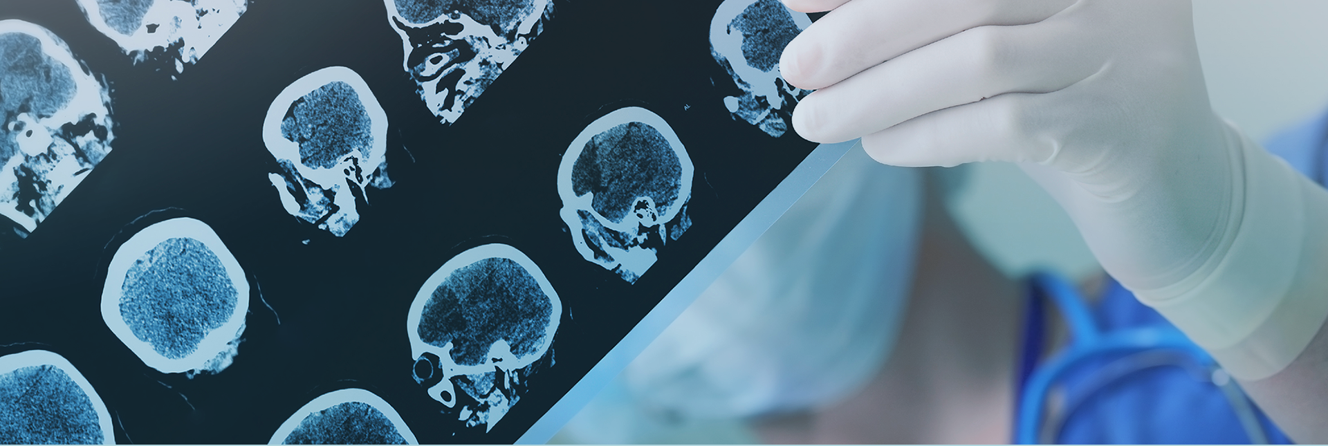
CT SCAN | COMPUTED TOMOGRAPHY SCAN
MRI | MAGNETIC RESONANCE IMAGING

Computed Axial Tomography (CT) and Magnetic Resonance Imaging (MRI) are important means of diagnosis – complementary to clinical examination and EEG – in the evaluation of a person with seizures. These neuroimaging techniques are especially sensitive to structural lesions of the central nervous system (CNS).
Magnetic resonance imaging uses a strong magnetic field and radio waves to create images of tissues, organs and other structures inside the body that are then viewed on a computer. This examination may reveal whether there is a structural cause for epilepsy. 
 
The MRI scan is not painful, but it can be very noisy, and some people feel very uncomfortable, having to stay in a limited space for some time. The radiology technician performing the examination may need to inject the person (in the hand or arm) with a harmless dye so that the tissues and blood vessels are better visualized in the image processed by the computer.
 
Some people develop allergies to this dye, and consequently the technician must ask the patient about his/her history of allergies. During the examination, the patient has to avoid movements and remain still, otherwise the scanned image may appear blurred and make it difficult for the doctor to interpret. 
 
Looking into the entire diagnosis phase, the doctor has several ways to collect and analyse information, including an accurate description of the suspected seizure event and the appropriate use of complementary tests, that are fundamental to a clear differentiation of epilepsy from other clinical conditions expressed also by seizures.